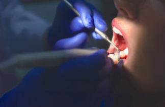
The diseases that present themselves in the oral cavity, the dental surgeon must be prepared to recognize them, because some alterations can be benign, malignant, or neoplasms that develop malignancy over time. There are several types of lesions that can occur in the mouth, and we must be careful to evaluate the oral cavity as a whole, each structure, so that if something is out of the ordinary, we can diagnose and refer it if necessary. And remember, whenever there is any doubt, to do complementary examinations.
- Leukoplakia: is a white lesion with the potential for malignant transformation, is not removed by scraping, and does not resemble any other lesion. It is more common in males, and can be found on the lower lip, tongue, lip commissure, and hard palate. Its diagnosis is confirmed by incisional biopsy followed by exfoliative cytology.
- Actinic cheilitis: it is a lesion due to UVA and UVB exposure with potential transformation into malignancy. It is more common in males, and is more common on the lower lip. The diagnosis is confirmed by incisional biopsy followed by exfoliative cytology.
- Candidiasis: popularly known as “thrush”, is a superficial fungal infection, observed more in immunosuppressed patients, and is removed by scraping. Exfoliative cytology biopsy can be done.
- Chronic atrophic candidiasis: Related to the upper total prosthesis, usually in elderly patients, carriers of ill-fitting prostheses for many years of use, traumatizing the mucosa. It has a velvety reddish coloration.
- Melanoma: this is an aggressive malignant neoplasm, which rarely affects the mouth, or appears late. It can affect the palate, the gingival mucosa, and the alveolar ridge.
- Erythroplakia: A reddish lesion, less common in the mouth, but potentially malignant. It affects the oral floor, hard or soft palate. It must be treated by the oncologist.
- Kaposi’s sarcoma: Human herpes virus (HHV-8). It is the first oral manifestation of AIDS, can develop into a malignant change, and its diagnosis is made with an incisional biopsy sent to histopathology.
- Epidermoid carcinoma: Malignant neoplasm of the oral cavity, which can be described as an ulcerated lesion, sharp elevated borders and hardened . It can be diagnosed by exfoliative cytology or histopathology.
- Papilloma: It is related to HPV, and the clinical appearance is characterized by a papule . It is located in the region of the uvula, soft or hard palate, and tongue, and less frequently on the mucosa. Removal of the lesion is recommended.
- Fibroma: It is a fibrous hyperplasia, recurrent to a trauma. It can be found on the jugal mucosa, the labial mucosa, or the lateral border of the tongue. Surgical removal is recommended, but the root cause must be eliminated to prevent recurrence.
- Hemangioma: This can be represented by a vascular malformation. When it occurs in children, it usually affects the skin or scalp and regresses spontaneously at puberty. To confirm the diagnosis, on-site vitro-pressure can be done. Treatment can be surgical for small lesions.
- Lipoma: It is a benign neoplasm of fatty tissue, its color is usually yellowish. It can be found on the jugal mucosa, lips, tongue, or oral floor. The treatment is surgical removal.
- Pyogenic Granuloma: Lesion resulting from an uncontrolled production of granulation tissue. It originates most often in the interdental papilla of the upper teeth and can also be found on the tongue, lips, oral mucosa or alveolar ridge.
Cysts
Cysts are common lesions, usually asymptomatic, but which can cause pain if infected, and can also cause deformity if large in proportion. True cysts are those that contain lined epithelium, containing fluid or semi-solid material. Those derived from remnants of materials from the process of odontogenesis are called odontogenic cysts. The pathological cavities not lined by epithelium are called pseudocysts. To obtain the diagnosis of the cyst, it should be referred for histopathology, and by the aspects of the capsule microscopically, the classification of the diagnosis of which cystic lesion is given.
Odontogenic:
- Periapical cyst: This is the most common of the odontogenic cysts, and originates from the epithelial remnants of malassez, and can develop due to the death of the pulp by caries, or by trauma. It is located in the apex of the tooth, and the treatment is surgical, by enucleation, and endodontic(root canal treatment) when the tooth is not compromised; when it is, tooth removal must be performed.
- Dentigerous cyst: Involves the crown of an unerupted tooth, most often on third molars and canines. Depending on the size, it can displace the tooth from its original position and adjacent teeth. The surgical treatment is removal by enucleation, if the third molar is involved, it is removed as well, but if it is canine, it is preferred to do a marsupialization for decompression of the cyst, to bring the involved tooth in its original position.
- Lateral periodontal cyst: Infrequent derived from the remains of the dental lamina (serres), preferably located in the mandible, in the region of the canines and pre molars, usually found at the apex of the most lateral roots. Treatment is surgical with enucleation.
Non Odontogenic
- Nasopalatine duct cyst: this originates inside the nasopalatine duct in the incisor canal, in the anterior midline. The treatment is surgical enucleation.
Pseudocysts
- Hemorrhagic cyst: This is not a true cyst, as it contains no epithelium; its etiology may have come from trauma. Treatment consists of filling the cavity with blood, to induce bone formation in the site; this can occur by surgical access or curettage.
- Aneurysmal bone cyst: This is an uncommon lesion, characterized by large spaces filled with blood, and separated by bands of fibrous tissue containing giant cells. It occurs preferentially in the posterior mandible, its behavior is generally aggressive, and its treatment is done by curettage, and in some cases there can be relapses.
Neoplasms
This group of pathology is very broad, with lesions of various complexities and variable behavior. Bone neoplasms are generally characterized by producing increased volume and deformity. The benign lesions grow more regularly and have more delimited radiographic aspects, while the malignant ones, on the contrary, rupture cortices, involve soft tissues, and demonstrate an infiltrative and diffuse aspect on radiographs.
- Ameloblastoma: This is a benign but invasive neoplasm that usually requires invasive and mutilating therapeutic interventions. It is most common in the molar and ramus region of the mandible. Its growth is slow, progressive, and painless, but very large lesions can present pain, paresthesia, and secondary infections. When the ameloblastoma presents unilocular, called unisystic, its treatment shows a better prognosis, even though it is a more conservative treatment. Those with multilocular forms known radiographically as “soap bubbles” are usually treated more aggressively with en bloc surgical resection. The dental surgeon must evaluate each case and each patient’s lesion histology, for the best planning and treatment.
- Myxoma: This is a slow, asymptomatic, locally invasive lesion, affecting the mandible more than the maxilla. Radiographically it is remembered as a “tennis racket” or “honeycomb”. The tumor can displace teeth or rupture cortical. The treatment is surgical removal of the tumor, but total removal is not always easy, and a larger resection may be required, with a good prognosis but with deformities in the region.
- Adenomatoid: It is located more in the maxilla, usually associated with unerupted canines; its growth is slow and asymptomatic. Treatment is done by curettage and generally extraction of the involved tooth, with no relapses and a good prognosis.
- Odontoma: These are common in both the mandible and maxilla. They are masses of dental tissues that compose themselves, resembling or not the dental morphology, generally associated in unerupted teeth. The treatment is surgical enucleation and conservative enucleation.
- Osteosarcoma: This is a malignant neoplasm in bone, with recurrence in the maxilla. The patient experiences symptoms such as pain, paresthesia, tooth mobility, and nasal obstruction. Radiographically the lesion is aggressive with bone destruction, and quickly it can reach adjacent soft tissues. This lesion can metastasize to other parts of the body, especially the lung. The recommended treatment is radical surgery with adjuvant chemotherapy.
- Lymphoma: These are malignant neoplasms derived from lymphocytes.
- Hodgkin type lymphoma: It begins in a cervical, inguinal, axillary lymph node. The disease can cause deformity as it progresses. The biopsy must be aspirated and treatment is by a combination of chemotherapy and radiotherapy.
- Non-Hodgkin type lymphoma: May start outside the lymph nodes, with a worse prognosis than the Hodgkin type, may be involved in people with HIV. It can present as ulcerated lesions or nodular expressions. Treatment is the same as for the Hodgkin type.

